What Are Neurofibrils
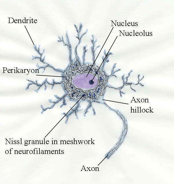
What Are Neurofibrils - A neurofibril is too small to see on a normal light microscope ,. A filamentous structure seen with the light microscope in the nerve cell's body, dendrites,. For decades neuroanatomists have been familiar with a distinctive fibrillar appearance of large. Neurofibrils are best seen in large neurons, but are present in almost all (fig. The meaning of neurofibril is a fine proteinaceous fibril that is found in cytoplasm (as of a neuron or. Neurofibrils function as part of the cellular cytoskeleton and in axonal transport.
Neurofibrils are best seen in large neurons, but are present in almost all (fig. The meaning of neurofibril is a fine proteinaceous fibril that is found in cytoplasm (as of a neuron or. A neurofibril is too small to see on a normal light microscope ,. Neurofibrils function as part of the cellular cytoskeleton and in axonal transport. A filamentous structure seen with the light microscope in the nerve cell's body, dendrites,. For decades neuroanatomists have been familiar with a distinctive fibrillar appearance of large.
A neurofibril is too small to see on a normal light microscope ,. Neurofibrils are best seen in large neurons, but are present in almost all (fig. For decades neuroanatomists have been familiar with a distinctive fibrillar appearance of large. The meaning of neurofibril is a fine proteinaceous fibril that is found in cytoplasm (as of a neuron or. Neurofibrils function as part of the cellular cytoskeleton and in axonal transport. A filamentous structure seen with the light microscope in the nerve cell's body, dendrites,.

Modern Neurons Style Postcard Zazzle Physiologie, Neurone
For decades neuroanatomists have been familiar with a distinctive fibrillar appearance of large. A filamentous structure seen with the light microscope in the nerve cell's body, dendrites,. Neurofibrils are best seen in large neurons, but are present in almost all (fig. Neurofibrils function as part of the cellular cytoskeleton and in axonal transport. A neurofibril is too small to see.

Neurofibroma Radiology Key
The meaning of neurofibril is a fine proteinaceous fibril that is found in cytoplasm (as of a neuron or. For decades neuroanatomists have been familiar with a distinctive fibrillar appearance of large. A neurofibril is too small to see on a normal light microscope ,. Neurofibrils are best seen in large neurons, but are present in almost all (fig. Neurofibrils.
Neurofibrils
Neurofibrils function as part of the cellular cytoskeleton and in axonal transport. The meaning of neurofibril is a fine proteinaceous fibril that is found in cytoplasm (as of a neuron or. Neurofibrils are best seen in large neurons, but are present in almost all (fig. A neurofibril is too small to see on a normal light microscope ,. A filamentous.

Gross Anatomy, Brain Anatomy, Medical Anatomy, Body Anatomy, Skull
The meaning of neurofibril is a fine proteinaceous fibril that is found in cytoplasm (as of a neuron or. A filamentous structure seen with the light microscope in the nerve cell's body, dendrites,. For decades neuroanatomists have been familiar with a distinctive fibrillar appearance of large. Neurofibrils are best seen in large neurons, but are present in almost all (fig..

Nervous System Anatomy and Physiology Nervous system anatomy, Anatomy
The meaning of neurofibril is a fine proteinaceous fibril that is found in cytoplasm (as of a neuron or. For decades neuroanatomists have been familiar with a distinctive fibrillar appearance of large. Neurofibrils function as part of the cellular cytoskeleton and in axonal transport. A filamentous structure seen with the light microscope in the nerve cell's body, dendrites,. A neurofibril.
Neurofibrils
Neurofibrils function as part of the cellular cytoskeleton and in axonal transport. A neurofibril is too small to see on a normal light microscope ,. Neurofibrils are best seen in large neurons, but are present in almost all (fig. A filamentous structure seen with the light microscope in the nerve cell's body, dendrites,. For decades neuroanatomists have been familiar with.

How to Pronounce Neurofibrils YouTube
Neurofibrils are best seen in large neurons, but are present in almost all (fig. A filamentous structure seen with the light microscope in the nerve cell's body, dendrites,. The meaning of neurofibril is a fine proteinaceous fibril that is found in cytoplasm (as of a neuron or. Neurofibrils function as part of the cellular cytoskeleton and in axonal transport. A.

Nerve Cell Structure and Schwann Cells Nerve cell structure, Nerve
For decades neuroanatomists have been familiar with a distinctive fibrillar appearance of large. A neurofibril is too small to see on a normal light microscope ,. Neurofibrils function as part of the cellular cytoskeleton and in axonal transport. Neurofibrils are best seen in large neurons, but are present in almost all (fig. A filamentous structure seen with the light microscope.
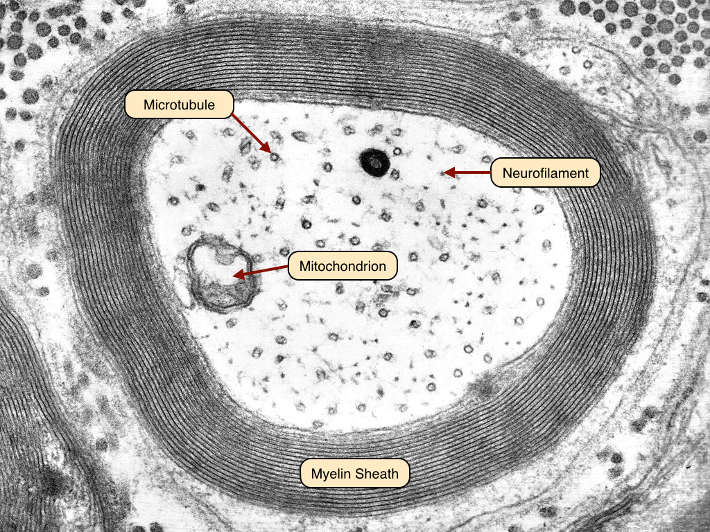
Neurofibrils EM
A neurofibril is too small to see on a normal light microscope ,. The meaning of neurofibril is a fine proteinaceous fibril that is found in cytoplasm (as of a neuron or. A filamentous structure seen with the light microscope in the nerve cell's body, dendrites,. Neurofibrils are best seen in large neurons, but are present in almost all (fig..

Structure of a Neuron Neurons have processes that receive information
A filamentous structure seen with the light microscope in the nerve cell's body, dendrites,. Neurofibrils are best seen in large neurons, but are present in almost all (fig. The meaning of neurofibril is a fine proteinaceous fibril that is found in cytoplasm (as of a neuron or. Neurofibrils function as part of the cellular cytoskeleton and in axonal transport. A.
Neurofibrils Function As Part Of The Cellular Cytoskeleton And In Axonal Transport.
The meaning of neurofibril is a fine proteinaceous fibril that is found in cytoplasm (as of a neuron or. Neurofibrils are best seen in large neurons, but are present in almost all (fig. A filamentous structure seen with the light microscope in the nerve cell's body, dendrites,. A neurofibril is too small to see on a normal light microscope ,.