What Is Opacification Of Left Mastoid Air Cells
What Is Opacification Of Left Mastoid Air Cells - I need to understand what it means if a ct scan of head shows opacification of the left mastoid air cells with most of these air cells sclerosed. Mastoid air cell opacification can occur in a number of situations and can include a spectrum of inflammatory, neoplastic, vascular,. What does opacification of the ethmoid air cells mean? Sinonasal inflammatory disease with sinus ostial. Opacification of mastoid air cells refers to the abnormal filling of these interconnected spaces within the mastoid bone. (a) transverse ct image through the left temporal bone shows complete opacification of the left tympanum and mastoid air cells.
Mastoid air cell opacification can occur in a number of situations and can include a spectrum of inflammatory, neoplastic, vascular,. Sinonasal inflammatory disease with sinus ostial. Opacification of mastoid air cells refers to the abnormal filling of these interconnected spaces within the mastoid bone. What does opacification of the ethmoid air cells mean? (a) transverse ct image through the left temporal bone shows complete opacification of the left tympanum and mastoid air cells. I need to understand what it means if a ct scan of head shows opacification of the left mastoid air cells with most of these air cells sclerosed.
I need to understand what it means if a ct scan of head shows opacification of the left mastoid air cells with most of these air cells sclerosed. Opacification of mastoid air cells refers to the abnormal filling of these interconnected spaces within the mastoid bone. Mastoid air cell opacification can occur in a number of situations and can include a spectrum of inflammatory, neoplastic, vascular,. Sinonasal inflammatory disease with sinus ostial. What does opacification of the ethmoid air cells mean? (a) transverse ct image through the left temporal bone shows complete opacification of the left tympanum and mastoid air cells.

Temporal Bone, Mastoid Air Cells Diagram Quizlet
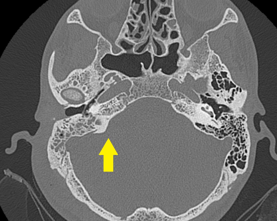
(a) transverse ct image through the left temporal bone shows complete opacification of the left tympanum and mastoid air cells. I need to understand what it means if a ct scan of head shows opacification of the left mastoid air cells with most of these air cells sclerosed. Mastoid air cell opacification can occur in a number of situations and.
Mastoid Air Cells Mri
I need to understand what it means if a ct scan of head shows opacification of the left mastoid air cells with most of these air cells sclerosed. Mastoid air cell opacification can occur in a number of situations and can include a spectrum of inflammatory, neoplastic, vascular,. Sinonasal inflammatory disease with sinus ostial. What does opacification of the ethmoid.

Comparison of CT findings (mastoid air cellstop & mandibular
Sinonasal inflammatory disease with sinus ostial. I need to understand what it means if a ct scan of head shows opacification of the left mastoid air cells with most of these air cells sclerosed. What does opacification of the ethmoid air cells mean? (a) transverse ct image through the left temporal bone shows complete opacification of the left tympanum and.
Mastoid Air Cells Anatomy
What does opacification of the ethmoid air cells mean? (a) transverse ct image through the left temporal bone shows complete opacification of the left tympanum and mastoid air cells. Sinonasal inflammatory disease with sinus ostial. Opacification of mastoid air cells refers to the abnormal filling of these interconnected spaces within the mastoid bone. I need to understand what it means.

Mastoid Air Cells Anatomy
Mastoid air cell opacification can occur in a number of situations and can include a spectrum of inflammatory, neoplastic, vascular,. What does opacification of the ethmoid air cells mean? Sinonasal inflammatory disease with sinus ostial. (a) transverse ct image through the left temporal bone shows complete opacification of the left tympanum and mastoid air cells. Opacification of mastoid air cells.

Mastoid Air Cells CrossSectional View Anatomy Diagram Quizlet
Mastoid air cell opacification can occur in a number of situations and can include a spectrum of inflammatory, neoplastic, vascular,. What does opacification of the ethmoid air cells mean? (a) transverse ct image through the left temporal bone shows complete opacification of the left tympanum and mastoid air cells. Sinonasal inflammatory disease with sinus ostial. Opacification of mastoid air cells.

Mastoid Air Cell Disease
I need to understand what it means if a ct scan of head shows opacification of the left mastoid air cells with most of these air cells sclerosed. Mastoid air cell opacification can occur in a number of situations and can include a spectrum of inflammatory, neoplastic, vascular,. What does opacification of the ethmoid air cells mean? (a) transverse ct.

Classification of the mastoid air cells (MACs). ‘a’ is the straight
(a) transverse ct image through the left temporal bone shows complete opacification of the left tympanum and mastoid air cells. What does opacification of the ethmoid air cells mean? I need to understand what it means if a ct scan of head shows opacification of the left mastoid air cells with most of these air cells sclerosed. Sinonasal inflammatory disease.

Mastoid Air Cells Anatomy
(a) transverse ct image through the left temporal bone shows complete opacification of the left tympanum and mastoid air cells. What does opacification of the ethmoid air cells mean? I need to understand what it means if a ct scan of head shows opacification of the left mastoid air cells with most of these air cells sclerosed. Opacification of mastoid.

Mastoid Air Cells Mri
I need to understand what it means if a ct scan of head shows opacification of the left mastoid air cells with most of these air cells sclerosed. Opacification of mastoid air cells refers to the abnormal filling of these interconnected spaces within the mastoid bone. What does opacification of the ethmoid air cells mean? Mastoid air cell opacification can.
What Does Opacification Of The Ethmoid Air Cells Mean?
Sinonasal inflammatory disease with sinus ostial. Mastoid air cell opacification can occur in a number of situations and can include a spectrum of inflammatory, neoplastic, vascular,. I need to understand what it means if a ct scan of head shows opacification of the left mastoid air cells with most of these air cells sclerosed. (a) transverse ct image through the left temporal bone shows complete opacification of the left tympanum and mastoid air cells.